Western Blot Knowledge Base A Complete Guide
Western blotting, also known as immunoblotting, is one of the most widely used techniques in molecular biology, biochemistry, and medical research. It allows scientists to detect, identify, and quantify specific proteins from complex biological samples. A solid understanding of the Western blot knowledge base is essential for both beginners and experienced researchers to obtain accurate and reproducible results. This guide covers the principles, materials, procedures, troubleshooting, and best practices associated with Western blotting.
1. Principles of Western Blotting
The core principle of Western blotting relies on specific antibody-antigen interactions. Proteins, which act as antigens, are immobilized on a membrane and detected using antibodies that specifically bind to the target protein. This allows researchers to determine not only the presence of a protein but also its approximate molecular weight and relative abundance.
The process combines two key techniques:
-
Protein separation using SDS-PAGE, which separates gel electrophoresis process based on size.
-
Protein detection using immunological methods, where antibodies recognize specific proteins.
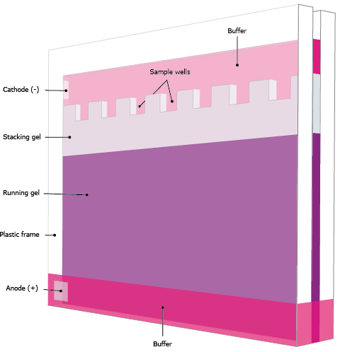
2. Essential Materials and Equipment
A Western blot requires several key materials:
-
Protein samples: Extracted from cells, tissues, or purified sources.
-
SDS-PAGE gels: For separating proteins by molecular weight.
-
Membranes: Nitrocellulose or PVDF membranes for immobilizing proteins.
-
Antibodies:
-
Primary antibodies bind to the protein of interest.
-
Secondary antibodies are enzyme-conjugated to amplify the signal.
-
-
Buffers and reagents: Running buffer, transfer buffer, blocking solution, wash buffers, and enzyme substrates.
-
Ladders or molecular weight markers: Used to estimate protein size.
-
Transfer apparatus: Either wet or semi-dry, depending on protein size and lab setup.
3. Step-by-Step Western Blot Procedure
Step 1: Sample Preparation
Proteins are extracted using lysis buffers containing detergents and protease inhibitors. The protein concentration is measured to ensure equal loading across samples. Samples are mixed with SDS-containing loading buffer and heat-denatured.
Step 2: SDS-PAGE
Proteins are separated on a polyacrylamide gel based on size. SDS ensures proteins carry a uniform negative charge, allowing size-based separation.
Step 3: Protein Transfer
Proteins are transferred from the gel to a membrane using a wet or semi-dry transfer apparatus. Wet transfer is ideal for large proteins and ensures uniform migration, while semi-dry transfer is faster and uses less buffer, suitable for small to medium proteins.
Step 4: Blocking
The membrane is incubated with a blocking solution (e.g., non-fat milk or BSA) to prevent nonspecific binding of antibodies.
Step 5: Antibody Incubation
The membrane is incubated with the primary antibody, followed by washing and incubation with an enzyme-conjugated secondary antibody. This amplifies the signal for detection.
Step 6: Detection
Detection is achieved using substrates that produce either light (chemiluminescent) or color (colorimetric). The intensity of the resulting bands correlates with protein abundance.
Step 7: Data Analysis
Protein bands are compared to molecular weight markers. Loading controls like β-actin or GAPDH ensure equal protein loading, and densitometry can quantify protein levels.
4. Common Issues and Troubleshooting
-
Weak or no signal: Could be due to low protein concentration, inefficient transfer, or improper antibody use.
-
High background: Often caused by insufficient blocking or excessive antibody concentration.
-
Multiple bands: May result from protein degradation or nonspecific antibody binding.
-
Uneven transfer or distorted bands: Could be caused by air bubbles or uneven gel placement.
Optimizing sample preparation, antibody concentrations, transfer conditions, and washing steps is critical for reliable results.
5. Applications of Western Blotting
Western blotting is widely applied in:
-
Protein expression analysis: Measuring changes in protein levels under different conditions.
-
Post-translational modifications: Detecting phosphorylation, ubiquitination, and glycosylation.
-
Recombinant protein verification: Confirming protein expression and size.
-
Medical research: Identifying biomarkers, studying disease mechanisms, and monitoring therapeutic effects.
Conclusion
The Western blot knowledge base encompasses the fundamental principles, essential materials, step-by-step procedures, troubleshooting strategies, and applications of this powerful technique. By mastering these aspects, researchers can accurately detect and quantify proteins, making Western blotting an indispensable tool in biological research. A thorough understanding of the method, coupled with careful experimental planning, ensures reproducibility, sensitivity, and specificity in protein analysis.